Home
/ Labeled Anatomy Of Rib Cage : Rib Anatomy at University of Medicine and Dentistry of New ... : In clinical applications, the sternal angle can be palpated at the t4 vertebral level.
Labeled Anatomy Of Rib Cage : Rib Anatomy at University of Medicine and Dentistry of New ... : In clinical applications, the sternal angle can be palpated at the t4 vertebral level.
Labeled Anatomy Of Rib Cage : Rib Anatomy at University of Medicine and Dentistry of New ... : In clinical applications, the sternal angle can be palpated at the t4 vertebral level.. The thoracic cavity is the more superior subdivision of the anterior cavity, and it is enclosed by the rib cage. It is sometimes called the lumbar region. These vertebrae form the foundation of the thoracic region's sturdy spinal column that supports the neck above, the rib cage, soft tissues, flexible joints, blood vessels, and nerves. The thoracic cavity contains the lungs and the heart, which is located in the mediastinum. Each are symmetrically paired on a right and left side.
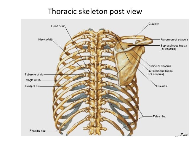
Each are symmetrically paired on a right and left side. The costal angle also marks the attachment for some of the deep back muscles to. It forms the bony framework for breathing. This is where the diaphragm separates the thoracic cavity from the abdominal cavity. The body, or shaft, of the rib is thin, flat and curved.
Thoracic cage from image.slidesharecdn.com The anterior (ventral) cavity has two main subdivisions: This point is marked with an x in the. The costal angle also marks the attachment for some of the deep back muscles to. Mar 20, 2015 · the human rib cage is made up of 12 paired rib bones; The thoracic cavity and the abdominopelvic cavity (see figure 4). Dog anatomy is not very difficult to understand if a labeled diagram is present to provide a graphic illustration of the same. The body, or shaft, of the rib is thin, flat and curved. Keep moving down until you feel the bottom edge of the rib cage;
Jul 08, 2021 · the best way to learn anatomy is to repeat as much as you can.
That is exactly what you will find in this dogappy article. The thoracic spine has 12 vertebrae stacked on top of each other, labeled from t1 down to t12. Nov 05, 2019 · related posts of rib cage diagram with organs anatomy of human stomach. Dog anatomy is not very difficult to understand if a labeled diagram is present to provide a graphic illustration of the same. The costal angle also marks the attachment for some of the deep back muscles to. It provides information about a dog's skeletal, reproductive, internal, and external anatomy, along with accompanying labeled diagrams. The thoracic cavity is the more superior subdivision of the anterior cavity, and it is enclosed by the rib cage. It is sometimes called the lumbar region. Use your fingers to probe the chest area of the pig. Jun 10, 2021 · the thoracic cage is a component of the thoracic wall and encloses the majority of the structures of the respiratory system. Keep moving down until you feel the bottom edge of the rib cage; The thoracic cavity and the abdominopelvic cavity (see figure 4). This is where the diaphragm separates the thoracic cavity from the abdominal cavity.
The anterior (ventral) cavity has two main subdivisions: Of all 24 ribs, the first seven pairs are often labeled as 'true.' Use your fingers to probe the chest area of the pig. The body, or shaft, of the rib is thin, flat and curved. The curve becomes most prominent at the costal angle, which is when the rib turns anterolaterally.
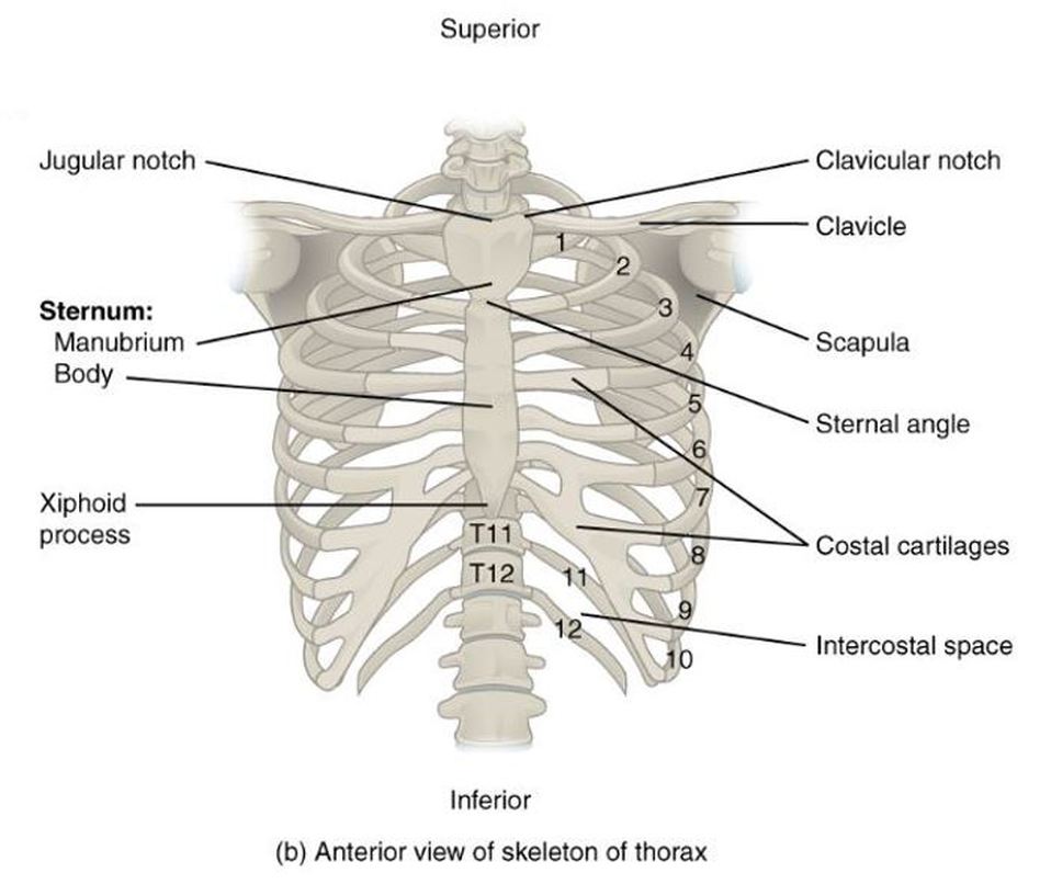
Manubrium sternal; Manubrium | Human ribs, Body anatomy ... from i.pinimg.com Keep moving down until you feel the bottom edge of the rib cage; Jun 10, 2021 · the thoracic cage is a component of the thoracic wall and encloses the majority of the structures of the respiratory system. Use your fingers to probe the chest area of the pig. The thoracic cavity contains the lungs and the heart, which is located in the mediastinum. You should be able to feel the hard sternum (breastbone) and the tiny ridges of the ribcage. The flank or latus is the side of the body between the rib cage and the iliac bone of the hip (below the rib cage and above the ilium). In clinical applications, the sternal angle can be palpated at the t4 vertebral level. The dome shaped thoracic cage provides the necessary rigidity for organ protection, weight support for the upper limbs and anchorage for muscles.
It forms the bony framework for breathing.
Quizzes are the secret to your success! The dome shaped thoracic cage provides the necessary rigidity for organ protection, weight support for the upper limbs and anchorage for muscles. Dog anatomy is not very difficult to understand if a labeled diagram is present to provide a graphic illustration of the same. This is where the diaphragm separates the thoracic cavity from the abdominal cavity. The costal angle also marks the attachment for some of the deep back muscles to. Jul 08, 2021 · the best way to learn anatomy is to repeat as much as you can. Use your fingers to probe the chest area of the pig. Of all 24 ribs, the first seven pairs are often labeled as 'true.' Anatomy of human stomach 10 photos of the anatomy of human stomach anatomy human colon, anatomy human digestive system, anatomy human heart, anatomy human kidney, anatomy human liver, anatomy human pancreas, anatomy human spleen, human body stomach, stomach, anatomy human colon, anatomy human digestive system, anatomy. The thoracic cavity contains the lungs and the heart, which is located in the mediastinum. Nov 05, 2019 · related posts of rib cage diagram with organs anatomy of human stomach. These vertebrae form the foundation of the thoracic region's sturdy spinal column that supports the neck above, the rib cage, soft tissues, flexible joints, blood vessels, and nerves. Jun 10, 2021 · the thoracic cage is a component of the thoracic wall and encloses the majority of the structures of the respiratory system.
It forms the bony framework for breathing. That is exactly what you will find in this dogappy article. The anterior (ventral) cavity has two main subdivisions: Nov 05, 2019 · related posts of rib cage diagram with organs anatomy of human stomach. It provides information about a dog's skeletal, reproductive, internal, and external anatomy, along with accompanying labeled diagrams.
the Thoracic Cage - SCIENTIST CINDY from www.scientistcindy.com The anterior (ventral) cavity has two main subdivisions: Each are symmetrically paired on a right and left side. It forms the bony framework for breathing. It is sometimes called the lumbar region. Of all 24 ribs, the first seven pairs are often labeled as 'true.' This is where the diaphragm separates the thoracic cavity from the abdominal cavity. The thoracic cavity contains the lungs and the heart, which is located in the mediastinum. You should be able to feel the hard sternum (breastbone) and the tiny ridges of the ribcage.
The body, or shaft, of the rib is thin, flat and curved.
You should be able to feel the hard sternum (breastbone) and the tiny ridges of the ribcage. It is sometimes called the lumbar region. Anatomy of human stomach 10 photos of the anatomy of human stomach anatomy human colon, anatomy human digestive system, anatomy human heart, anatomy human kidney, anatomy human liver, anatomy human pancreas, anatomy human spleen, human body stomach, stomach, anatomy human colon, anatomy human digestive system, anatomy. In clinical applications, the sternal angle can be palpated at the t4 vertebral level. The sternal angle, which varies around 162 degrees in males, marks the approximate level of the 2nd pair of costal cartilages, which attach to the second ribs, and the level of the intervertebral disc between t4 and t5. Each are symmetrically paired on a right and left side. The thoracic cavity contains the lungs and the heart, which is located in the mediastinum. The thoracic spine has 12 vertebrae stacked on top of each other, labeled from t1 down to t12. Nov 05, 2019 · related posts of rib cage diagram with organs anatomy of human stomach. It provides information about a dog's skeletal, reproductive, internal, and external anatomy, along with accompanying labeled diagrams. The dome shaped thoracic cage provides the necessary rigidity for organ protection, weight support for the upper limbs and anchorage for muscles. The flank or latus is the side of the body between the rib cage and the iliac bone of the hip (below the rib cage and above the ilium). The costal angle also marks the attachment for some of the deep back muscles to.
Each are symmetrically paired on a right and left side anatomy of rib cage. The thoracic cavity is the more superior subdivision of the anterior cavity, and it is enclosed by the rib cage.
